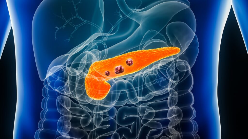
A team of researchers at MIT’s CSAIL division, specializing in computer engineering and AI development, have developed a unique neural network named “PRISM”. This network, built on two machine learning algorithms, is designed to detect pancreatic ductal adenocarcinoma (PDAC), the most common type of pancreatic cancer, with a higher accuracy than existing diagnostic standards.
The current PDAC screening criteria only detect about 10% of cases when examined by professionals. In contrast, PRISM has shown a significant improvement, identifying PDAC cases 35% of the time.
What is the Accuracy of PRISM?
PRISM, a neural network developed by the CSAIL division at MIT, has made significant strides in the detection of pancreatic ductal adenocarcinoma (PDAC). While existing PDAC screening criteria can only detect about 10% of cases, PRISM has demonstrated the ability to identify PDAC cases 35% of the time.
In a simulated model deployment, PRISM’s sensitivity was 35.9% with a specificity of 95.3% at a Standardised Incidence Ratio (SIR) of 5.10, which is higher than the current screening inclusion threshold. This indicates that PRISM could detect 3.5 times more cases at a similar risk level compared to current screening guidelines. These findings underscore the potential of AI in enhancing the accuracy and early detection of pancreatic cancer.
What sets PRISM apart is its development process. The neural network was trained using a diverse range of real electronic health records from various health institutions across the US. Over 5 million patient records were used, a scale of data that surpasses what is typically used in this area of research. The model uses routine clinical and lab data to make predictions, offering a significant advancement over other PDAC models, which are often limited to specific geographic regions.
The PRISM project, which began over six years ago, was motivated by the need for early detection of PDAC. This is crucial as most patients are diagnosed in the later stages of the cancer’s development, with about eighty percent being diagnosed far too late.
PRISM works by analyzing various factors such as patient demographics, previous diagnoses, current and previous medications in care plans, and lab results. It predicts the probability of cancer by analyzing electronic health record data along with factors like a patient’s age and certain lifestyle risk factors.
However, PRISM’s ability to help diagnose is currently limited by the rate at which the AI can reach the masses. The technology is currently confined to MIT labs and select patients in the US. To increase accessibility, the AI will need to be fed more diverse data sets and perhaps even global health profiles.
This is not MIT’s first attempt at developing an AI model that can predict cancer risk. They have previously trained models to predict the risk of breast cancer among women using mammogram records. In this line of research, MIT experts confirmed that the more diverse the data sets, the better the AI gets at diagnosing cancers across diverse races and populations.
The continued development of AI models that can predict cancer probability will not only improve outcomes for patients if malignancy is identified earlier, but it will also lessen the workload of overworked medical professionals. The potential for change in the diagnostics market is so significant that it has attracted the interest of big tech commercial companies like IBM, which has attempted to create an AI program that can detect breast cancer a year in advance.
The development and application of AI in the field of medical diagnostics, particularly in cancer detection, is a rapidly evolving field. With advancements like PRISM, we are moving closer to a future where early and accurate diagnosis could significantly improve patient outcomes.
Artificial Intelligence in the Detection of Other Cancers
AI has been increasingly used in the detection of various types of cancer. Here are a few examples:
Lung Cancer: An AI tool developed by experts at the Royal Marsden NHS Foundation Trust, the Institute of Cancer Research, London, and Imperial College London can identify whether abnormal growths found on CT scans are cancerous1. The algorithm performs more efficiently and effectively than current methods1.
Multiple Tissue Types: Paige, a global leader in end-to-end digital pathology solutions and clinical AI applications, has developed an application that can detect cancer across more than 17 different tissue types including skin, lung, and gastrointestinal tract, along with multiple rare tumor types and metastatic deposits2.
Various Cancers: A deep-learning computer program was able to detect the presence of molecular and genetic alterations based only on tumor images across multiple cancer types, including head and neck cancer3. AI contributes significantly to cancer detection by emulating the human brain’s information processing. In cancer diagnosis, AI analyses radiological and pathological images, learning from extensive datasets to recognize unique features associated with various cancers4.
These advancements in AI technology are revolutionizing the field of cancer diagnostics, enabling earlier detection and treatment, and potentially improving patient outcomes.
Naorem Mohen is the Editor of Signpost News. Explore his views and opinion on X: @laimacha.

